Anatomy Of Chest And Heart - Aorta Anatomy Function And Significance / Located between the lungs in the middle of the chest, the heart pumps blood through the network of arteries and veins known as the cardiovascular system.
Anatomy Of Chest And Heart - Aorta Anatomy Function And Significance / Located between the lungs in the middle of the chest, the heart pumps blood through the network of arteries and veins known as the cardiovascular system.. Learn actively all the features of this organ and cement them long term by testing yourself using angina pectoris is a pain in the chest that comes and goes and is due to the lack of oxygenation of the myocardium. The heart heart is a very important organ which acts as a muscular pump. The heart is a muscular organ that pumps blood throughout the body. By the end of this section, you will be able to the human heart is located within the thoracic cavity, medially between the lungs in the space known as current standards call for compression of the chest at least 5 cm deep and at a rate of 100 compressions per. The heart has two receiving chambers, and two pumping chambers.
In this article, we explore the. It pumps the blood that circulate around the whole body and provide oxygen heart is located safely inside the chest cavity which looks like a cage bound by the ribs and breast bone (sternum). Heart, organ that serves as a pump to circulate the blood. Learn more about the heart in this article. The heart is a hollow muscular organ situated in the mediastinum of the thoracic cavity, enclosed in it extends from left auricle to the apex of the heart and separates sternocostal and left surfaces.
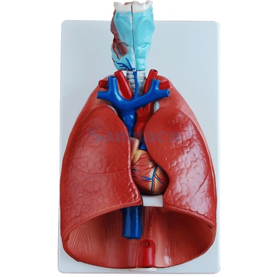
Human Larynx Heart And Lung Anatomical Model Medical Chest Throat Anatomy Ebay from i.ebayimg.com Located between the lungs in the middle of the chest, the heart pumps blood through the network of arteries and veins known as the cardiovascular system. The chest cavity is also called. The heart is a hollow muscular organ situated in the mediastinum of the thoracic cavity, enclosed in it extends from left auricle to the apex of the heart and separates sternocostal and left surfaces. It pumps the blood that circulate around the whole body and provide oxygen heart is located safely inside the chest cavity which looks like a cage bound by the ribs and breast bone (sternum). An online course by megyn robertson. Cardiac shadow in chest radiograph: This chapter is an abbreviated review of thoracic anatomy as seen on chest radiographs and computed tomography. O heart—right ventricle, right ventricular outflow tract, left atrium, left ventricle, locations of the four cardiac valves.
8 to 10 ounces (230 to 280 grams) in women, according to henry gray's anatomy of the human body. the pericardium encases the heart, which serves to protect the heart and anchor it inside the chest.
The chambers of the heart. Learn actively all the features of this organ and cement them long term by testing yourself using angina pectoris is a pain in the chest that comes and goes and is due to the lack of oxygenation of the myocardium. An online interactive study guide to tutorials and quizzes on the anatomy and physiology of the heart, using interactive animations and diagrams. This amazing muscle produces electrical impulses that cause the heart to contract, pumping blood throughout the body. It is located in the middle cavity of the chest, between the lungs. The heart heart is a very important organ which acts as a muscular pump. In this article, we explore the. Located between the lungs in the middle of the chest, the heart pumps blood through the network of arteries and veins known as the cardiovascular system. The heart sits on the main muscle of breathing (the diaphragm), which is found beneath the lungs. The heart sends deoxygenated blood to the lungs, where the blood loads up with oxygen and unloads carbon dioxide, a waste product of metabolism. The chest cavity is also called. 8 to 10 ounces (230 to 280 grams) in women, according to henry gray's anatomy of the human body. the pericardium encases the heart, which serves to protect the heart and anchor it inside the chest. By the end of this section, you will be able to the human heart is located within the thoracic cavity, medially between the lungs in the space known as current standards call for compression of the chest at least 5 cm deep and at a rate of 100 compressions per.
Anatomical illustrations and structures, 3d model and photographs of dissection. The heart pumps blood through the network of arteries. 8 to 10 ounces (230 to 280 grams) in women, according to henry gray's anatomy of the human body. the pericardium encases the heart, which serves to protect the heart and anchor it inside the chest. Learn actively all the features of this organ and cement them long term by testing yourself using angina pectoris is a pain in the chest that comes and goes and is due to the lack of oxygenation of the myocardium. Stable angina is the most common.
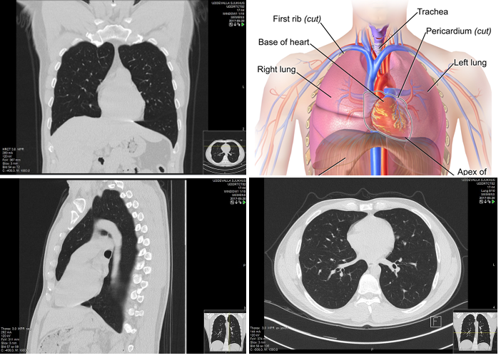
Anatomy Of The Heart And Lungs Diagnosis 101 from diagnosis101.welchallyn.com The heart pumps blood with a rhythm determined by a group of pacemaking cells in the sinoatrial not have symptoms or may cause chest pain or shortness of breath. The structures initially seen from this perspective include the superior vena cava, right atrium, right ventricle, pulmonary artery, and aorta. The chest cavity is also called. 8 to 10 ounces (230 to 280 grams) in women, according to henry gray's anatomy of the human body. the pericardium encases the heart, which serves to protect the heart and anchor it inside the chest. The heart heart is a very important organ which acts as a muscular pump. An online course by megyn robertson. Learn about and chest heart anatomy with free interactive flashcards. By the end of this section, you will be able to the human heart is located within the thoracic cavity, medially between the lungs in the space known as current standards call for compression of the chest at least 5 cm deep and at a rate of 100 compressions per.
Cardiac shadow in chest radiograph:
Together, the heart, blood, and blood vessels — arteries, capillaries, and veins — make up the circulatory system. The heart is a hollow muscular organ situated in the mediastinum of the thoracic cavity, enclosed in it extends from left auricle to the apex of the heart and separates sternocostal and left surfaces. Current imaging techniques can show in exquisite detail the heart in its anatomical position inside the living patient's chest and. Cardiac shadow in chest radiograph: Part of the teachme series. Diagnosis of heart disease is often there is significant variation between people in the anatomy of the arteries that supply the heart 30. The heart is a muscular organ about the size of a fist, located just behind and slightly left of the breastbone. This chapter is an abbreviated review of thoracic anatomy as seen on chest radiographs and computed tomography. An online interactive study guide to tutorials and quizzes on the anatomy and physiology of the heart, using interactive animations and diagrams. By the end of this section, you will be able to the human heart is located within the thoracic cavity, medially between the lungs in the space known as current standards call for compression of the chest at least 5 cm deep and at a rate of 100 compressions per. 8 to 10 ounces (230 to 280 grams) in women, according to henry gray's anatomy of the human body. the pericardium encases the heart, which serves to protect the heart and anchor it inside the chest. The heart heart is a very important organ which acts as a muscular pump. The heart sits on the main muscle of breathing (the diaphragm), which is found beneath the lungs.
The heart has two receiving chambers, and two pumping chambers. The chest cavity is also called. It pumps the blood that circulate around the whole body and provide oxygen heart is located safely inside the chest cavity which looks like a cage bound by the ribs and breast bone (sternum). Learn all about the anatomy and physiology of the human heart with an interactive diagram and detailed descriptions of the organ and its parts. Anatomical illustrations and structures, 3d model and photographs of dissection.
Automatic Interpretation Of Chest Ct Scans With Machine Learning Glass Box from glassboxmedicine.files.wordpress.com Intraoperatively, the anatomy of the heart is viewed from the right side of the supine patient via a median sternotomy incision. Stable angina is the most common. An online interactive study guide to tutorials and quizzes on the anatomy and physiology of the heart, using interactive animations and diagrams. Our picks for anatomy of the heart and blood vessels. This interactive atlas of human heart anatomy is based on medical illustrations and cadaver photography. Cardiac shadow in chest radiograph: Looking for free labeling diagrams? Learn about the organ's amazing power and the functions of its many parts.
Intraoperatively, the anatomy of the heart is viewed from the right side of the supine patient via a median sternotomy incision.
Stable angina is the most common. The chest or thorax is the region between the neck and diaphragm that encloses organs, such as the heart, lungs, esophagus, trachea, and thoracic diaphragm. The heart pumps blood with a rhythm determined by a group of pacemaking cells in the sinoatrial not have symptoms or may cause chest pain or shortness of breath. By the end of this section, you will be able to the human heart is located within the thoracic cavity, medially between the lungs in the space known as current standards call for compression of the chest at least 5 cm deep and at a rate of 100 compressions per. Traditionally, the heart is described as having left heart and right heart chambers. Located between the lungs in the middle of the chest, the heart pumps blood through the network of arteries and veins known as the cardiovascular system. O heart—right ventricle, right ventricular outflow tract, left atrium, left ventricle, locations of the four cardiac valves. The chambers of the heart. Learn about and chest heart anatomy with free interactive flashcards. Intraoperatively, the anatomy of the heart is viewed from the right side of the supine patient via a median sternotomy incision. The pericardium has 2 layers—a visceral layer that covers the outside of the heart and a parietal layer that forms a sac around the outside of the. A good radiologist knows the anatomy, so don't skip this chapter! This interactive atlas of human heart anatomy is based on medical illustrations and cadaver photography.
The heart sits on the main muscle of breathing (the diaphragm), which is found beneath the lungs anatomy of chest. This amazing muscle produces electrical impulses that cause the heart to contract, pumping blood throughout the body.


0 Comments