Foot Muscles Mri - Soleus Muscle Radiology Reference Article Radiopaedia Org / However, on mri images, no muscular abnormalities were detected.. There are 10 intrinsic muscles located in the sole of the foot. Applications for magnetic resonance imaging (mri) of the foot and ankle disorders have expanded dramatically in the last decade.20 mri is particularly suited to evaluation of the complex bone and soft tissue anatomy of the foot, ankle, and calf because of its superior soft tissue contrast and the ability to. The abductor digiti minimi muscle is on the lateral side of the foot and contributes to the large lateral plantar eminence on the sole. This means that the little toe can only be extended by the extensor digitorum longus muscle only. Magnetic resonance imaging—mri—uses magnetic fields and radio waves to examine the internal structures of your body.
Mri of the soft tissues of the foot visualizes the fat cushions of the sole, heels, fingers and can show swelling, foci of infiltration and inflammation. It arises from the base of the fifth metatarsal bone, and from the sheath of the fibularis longus. The muscles acting on the foot can be divided into two distinct groups; Involved early gray = muscle: Indications for foot mri scan.
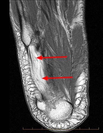
Baxter S Nerve First Branch Of The Lateral Plantar Nerve Impingement Radsource from radsource.us It must be placed in the center of the magnet, to obtain homogeneous fat. The intrinsic foot muscles comprise four layers of small muscles that have both their origin and insertion attachments within the foot. Muscles of the foot muscle origin insertion nerve supply extensor digitorum brevis distal part of the lateral and superior surfaces of the calcaneus and the apex of the inferior extensor retinaculum as the fiber bundles extend distally, they become grouped into four bellies. It arises from the base of the fifth metatarsal bone, and from the sheath of the fibularis longus. Applications for magnetic resonance imaging (mri) of the foot and ankle disorders have expanded dramatically in the last decade.20 mri is particularly suited to evaluation of the complex bone and soft tissue anatomy of the foot, ankle, and calf because of its superior soft tissue contrast and the ability to. Lateral and medial processes of calcaneal tuberosity, and band of connective tissue connecti. The flexor digiti minimi brevis (flexor brevis minimi digiti, flexor digiti quinti brevis) lies under the metatarsal bone on the little toe, and resembles one of the interossei. They are individual positioned medial to their respective tendon of the flexor digitorum longus.
The intrinsic foot muscles (ifm) are the main local stabilizers of the foot and are part of the active and neural subsystems that constitute the foot core.
Indications for foot mri scan. The difference in 18ffdg uptake between the patients and the controls was significant in muscle (p. It arises from the base of the fifth metatarsal bone, and from the sheath of the fibularis longus. Perform routine foot plus coronal fmpspgr fat saturated pre and post gad images and axial post gad fmpspgr fat saturated images. Epidemiology of tuberculosis etiology tuberculous spondylodiscitis clinical manifestations review of imaging findings: The abductor digiti minimi muscle is on the lateral side of the foot and contributes to the large lateral plantar eminence on the sole. Muscles of the foot are located on its rear and on the sole. This means that the little toe can only be extended by the extensor digitorum longus muscle only. Synovitis, tenosynovitis, bursitis, and ganglion cysts) > congenital and developmental conditions( eg.dysplasia, tarsal coalition). 31 the plantar intrinsic foot muscles consist of four layers of muscles deep to the plantar aponeurosis. The muscle that removes the big toe (m.abductor hallucis) lies superficially along the medial edge of the foot. Involved early gray = muscle: As a result, during walking the body's center of gravity normally fluctuates only 5cm in both vertical and lateral directions.
Top suggestions for foot muscle anatomy mri. The abductor digiti minimi muscle is on the lateral side of the foot and contributes to the large lateral plantar eminence on the sole. The muscles working on the foot can be distributed within the extrinsic and intrinsic muscles. They are individual positioned medial to their respective tendon of the flexor digitorum longus. It must be placed in the center of the magnet, to obtain homogeneous fat.
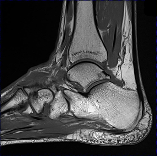
Ankle Tendons Topographic Anatomy Radiology Case Radiopaedia Org from prod-images-static.radiopaedia.org It arises from the base of the fifth metatarsal bone, and from the sheath of the fibularis longus. There can't be any metal in the room, not just where you have the mri. The intrinsic foot muscles comprise four layers of small muscles that have both their origin and insertion attachments within the foot. Involved early gray = muscle: Perform routine foot plus coronal fmpspgr fat saturated pre and post gad images and axial post gad fmpspgr fat saturated images. 12 photos of the foot muscle anatomy mri. Indications for foot mri scan. The intrinsic foot muscles (ifm) are the main local stabilizers of the foot and are part of the active and neural subsystems that constitute the foot core.
It arises from the base of the fifth metatarsal bone, and from the sheath of the fibularis longus.
Muscles of the foot are located on its rear and on the sole. In addition, an image of all the muscles of the back and plantar part of the foot, all tendons and tendon ligaments, blood vessels and nerves are obtained. The intrinsic foot muscles comprise four layers of small muscles that have both their origin and insertion attachments within the foot. Musculoskeletal system | muscle structure and function. Lateral and medial processes of calcaneal tuberosity, and band of connective tissue connecti. The purpose of this study was to investigate the relationship of muscle mri findings and gait disturbance in myotonic dystrophy type 1 (dm1) patients. Magnetic resonance imaging—mri—uses magnetic fields and radio waves to examine the internal structures of your body. Involved early gray = muscle: The muscle that removes the big toe (m.abductor hallucis) lies superficially along the medial edge of the foot. Mri with hardware in foot? There are 10 intrinsic muscles located in the sole of the foot. This means that the little toe can only be extended by the extensor digitorum longus muscle only. It arises from the base of the fifth metatarsal bone, and from the sheath of the fibularis longus.
There are 10 intrinsic muscles located in the sole of the foot. Interestingly the dorsal foot muscles generally have no insertion at the little toe. More runners in the minimalist shoe group had increases in bone marrow edema than in the control. The flexor digiti minimi brevis (flexor brevis minimi digiti, flexor digiti quinti brevis) lies under the metatarsal bone on the little toe, and resembles one of the interossei. Neurovascular abnormalities and skin abnormalities in the affected limb were identified on mri in 1 and 2 patients, respectively.
Mri Lower Extremities Leg Cedars Sinai from www.cedars-sinai.org Where you get the potential for problems with. Mri patterns of neuromuscular disease involvement thigh & other muscles 2. Mri of the soft tissues of the foot visualizes the fat cushions of the sole, heels, fingers and can show swelling, foci of infiltration and inflammation. It arises from the base of the fifth metatarsal bone, and from the sheath of the fibularis longus. Mri with hardware in foot? Interestingly the dorsal foot muscles generally have no insertion at the little toe. Epidemiology of tuberculosis etiology tuberculous spondylodiscitis clinical manifestations review of imaging findings: The muscle that removes the big toe (m.abductor hallucis) lies superficially along the medial edge of the foot.
The purpose of this study was to investigate the relationship of muscle mri findings and gait disturbance in myotonic dystrophy type 1 (dm1) patients.
There are 10 intrinsic muscles located in the sole of the foot. The muscle that removes the big toe (m.abductor hallucis) lies superficially along the medial edge of the foot. The difference in 18ffdg uptake between the patients and the controls was significant in muscle (p. The muscles acting on the foot can be divided into two distinct groups; The intrinsic foot muscles (ifm) are the main local stabilizers of the foot and are part of the active and neural subsystems that constitute the foot core. In addition, an image of all the muscles of the back and plantar part of the foot, all tendons and tendon ligaments, blood vessels and nerves are obtained. This article reviews the use of magnetic resonance imaging (mri) in the evaluation of the foot, including a discussion of bone and cartilage abnormalities in an article published in the august 2006 issue of this journal, the authors reviewed magnetic resonance imaging (mri) of the ankle. Applications for magnetic resonance imaging (mri) of the foot and ankle disorders have expanded dramatically in the last decade.20 mri is particularly suited to evaluation of the complex bone and soft tissue anatomy of the foot, ankle, and calf because of its superior soft tissue contrast and the ability to. Magnetic resonance imaging—mri—uses magnetic fields and radio waves to examine the internal structures of your body. The purpose of this study was to investigate the relationship of muscle mri findings and gait disturbance in myotonic dystrophy type 1 (dm1) patients. Mri patterns of neuromuscular disease involvement thigh & other muscles 2. Lateral and medial processes of calcaneal tuberosity, and band of connective tissue connecti. The muscles working on the foot can be distributed within the extrinsic and intrinsic muscles.


0 Comments